

Joe and I have had numerous people contact us wanting to see his brain scans to "see" the problem he is having. Herein lies the problem; unless one knows exactly what they are looking at/for it is hard to notice any abnormalities. Thus, this post was born. :)
Here is a comparison of a "normal" brain vs one of someone with hydrocephalus. Note the enlarged blue area, that is where the excess cerebrospinal fluid, or CSF, builds up within the ventricles.

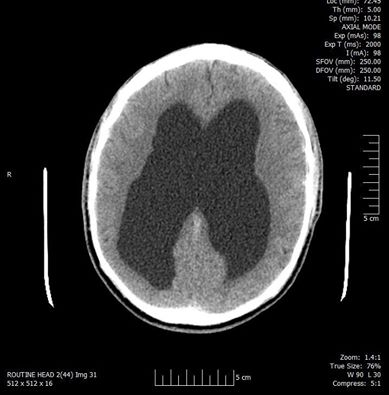
This little jem below is from the CT scan performed on Joe on March 4th, 2016. Note the dark/black portion, that is where the CSF has built up.
Now, since we are totally sharing EVERY aspect of Joe's hydrocephalus he has given me permission to share a cell phone recording of his actual CT scans:
Pretty neat looking if you ask us...well that's before we knew exactly what we were looking at. Wondering what caused this problem with Joe? Here is where things get interesting, well if you nerd out to science and medical related things anyway. We all have something called a cerebral aqueduct which is a narrow opening in the brain that connects the third ventricle with the fourth, allowing cerebrospinal fluid to flow between the two areas. The ventricles are the openings in the brain that provide a pathway for the CSF.

The cerebral aqueduct is about 3/4 of an inch long and is lined with ependyma which is a membrane that lines the ventricles of the brain. Unfortunately, Joe's aqueduct is stenosed or narrow which has created a blockage. Cerebrospinal fluid from the lateral ventricles, as well as the third, can now longer flow down to the fourth. So the fluid has not choice but to "back up" with no where to go. The goal is to create a new wider opening with surgery next month. If all goes well that should pretty much fix the issue.
No comments:
Post a Comment